Oncology
(O15) Innovative Development of Organoids from Canine Mastocytomas: A Novel In Vitro Model Background
Thursday, June 19, 2025
4:10 PM - 4:25 PM ET
Location: Exhibit Hall Poster Park - Kiosk 2

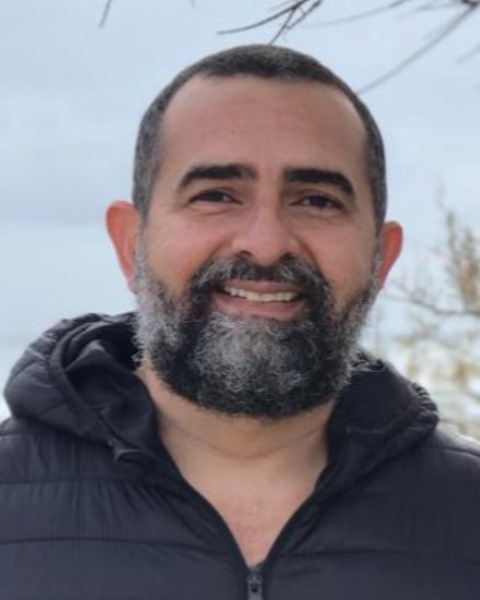
Andre Meneses, DVM, PhD (he/him/his)
Auxiliar Professor
Faculty of Veterinary Medicine, Lusofona University of Lisbon
LISBOA, Lisboa, Portugal
Research Abstract - ePoster Presenter(s)
Abstract: Canine mastocytomas are the most common cutaneous malignancy in dogs, providing a valuable model for studying mast cell tumor biology. Organoid technology has revolutionized cancer research by creating in vitro models that recapitulate tumor behavior and microenvironments. However, descriptions of organoids from canine mastocytomas are notably absent. This study addresses this gap by isolating and developing a specialized culture medium to sustain those organoids growth and morphology. The objective was to establish a reliable protocol for isolating these organoids and to develop a culture medium that supports their rapid growth and structural integrity. Tumor samples were obtained from three dogs with mastocytomas confirmed by histopathology. Ethical guidelines were followed, and informed owner consent was obtained. Tumor tissues were enzymatically and mechanically dissociated into single-cell suspensions and embedded in a 3D extracellular matrix. Organoids were cultured in a medium composed of Advanced DMEM/F12 supplemented with B27, Glutamax, N2, Hepes, R-spondin, Wnt-CM, FGF, NOG, EGF, A83, Y-27632, and SB202190. Growth was monitored daily, focusing on time to visible formation, confluence, and morphology. Organoids were visible 48 hours post-culture initiation in all three samples. The tailored medium supported rapid growth, reaching high confluence within 7 days. Morphological assessments revealed that organoids exhibited spheroidal structures and dense cellular clusters under phase-contrast microscopy. This study represents a pioneering effort to establish organoids from canine mastocytomas, highlighting the novelty and significance of this work, which provides a foundation for future investigations into tumor biology, drug discovery, and personalized treatment approaches in both veterinary and comparative oncology.


